Calcaneal fracture
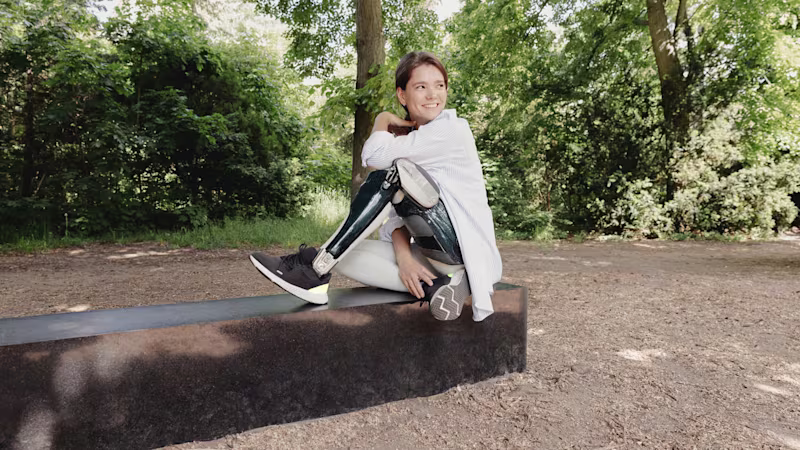
A calcaneal (heel bone) fracture is often caused by jumping from a great height. Orthoses can relieve the foot after an operation or may be used in conservative treatment.
Causes, symptoms and treatment
A heel bone fracture is often caused by jumping or falling from a great height. Men are affected five to eight times more frequently than women, and walking is restricted due to pain directly after the accident. In some cases, calcaneal fractures can be treated conservatively.
Ottobock supports and orthoses
Causes
A calcaneal fracture is generally caused by a high amount of force being exerted in the area of the heel bone. The most common cause for a calcaneal fracture is falling from a great height (approx. 80%), followed by traffic accidents (approx. 10%), sports accidents (approx. 8%) and fatigue fractures (2%). Bilateral fractures occur in 20 percent of all cases. Osteoporosis is also a possible causes, in which case a calcaneal fracture is an after-effect. Occupational and travel accidents account for half of all calcaneal fractures. Men are affected five to eight times more often than women, mainly between 35 and 60 years of age.
Symptoms
With a calcaneal fracture, acute swelling occurs in the anterior region and above the ankle joint. Most of the time, the entire foot is affected by the swelling, and considerable discolouration due to a haematoma is normally observed. Mobility of the foot is limited by severe pain.
In some cases, the skin and soft tissue in the area of the calcaneus tear during the accident. This results in a compound fracture with an increased risk of infection. Since this is an emergency, it requires immediate medical treatment in the form of an operation.
Diagnosis
Once the doctor has gained an initial impression based on a detailed medical history and a description of the accident, additional imaging procedures are used to make a diagnosis. X-rays are taken in three planes and an MRI (magnetic resonance imaging) is carried out. Computer tomography (CT) is required to classify the calcaneal fracture and plan the possible surgical intervention.
Therapy
If the calcaneal fracture is not displaced and no articular surfaces are affected, conservative treatment is usually recommended. Orthoses that relieve strain help speed up healing.
An acute calcaneal fracture is treated using the RICE method (rest, ice, compression, elevation). This is complemented by pain relievers and active physical therapy in the form of lymphatic drainage. The aim is to reduce the pronounced swelling and improve mobility in the tarsal bones where possible. Weight on the heel should be relieved for at least 6 to 12 weeks, depending on the progression.
In many cases, however, a calcaneal fracture is much more complex. Operations are performed on all calcaneal fractures involving the articular surfaces and with an offset of more than 1 mm in width. Surgery is also performed if the hindfoot is malpositioned. Generally, a right-angled incision is made on the outside of the calcaneus to operate in the affected area. The operation is usually carried out between 6 and 10 days after the accident due to the significant swelling in the soft tissue. Swelling in the affected foot should decrease in the meantime with the help of elevation, cooling and lymphatic drainage.
In the case of an open fracture, the destroyed tissue is surgically removed and the joint is fixed, usually by means of external connecting rods. During the operation, the bone fragments are initially assembled, the position of the calcaneus is improved, and it is returned to the correct position. Once the length and shape of the calcaneus have been restored, a plate with screws is used to stabilize it.
After the operation, the leg is initially supported in a padded splint until physiotherapy begins after two to five days. Weight bearing is usually avoided for at least eight to 12 weeks depending on the extent of the fracture and the stability obtained through surgery. Extreme strain on the heel should be avoided for the next six months, but the level of activity can be increased over time. X-rays are usually taken again after around six weeks, 12 weeks, six months and a year. A special heel relief orthosis can significantly improve mobility in the early phase.
Back to everyday activities: three steps to an Ottobock orthotic
- Here you’ll find an overview of all the orthotics and supports that could potentially help you. Take the list with you to your next doctor’s appointment.
- Talk to your doctor about which orthotic is best suited to your symptoms and condition. Your doctor can then write you a prescription for the appropriate orthotic.
- Take your prescription to a medical supply company. They’ll give you your new orthotic and adjust it to fit your exact body measurements.